Summary information and primary citation
- PDB-id
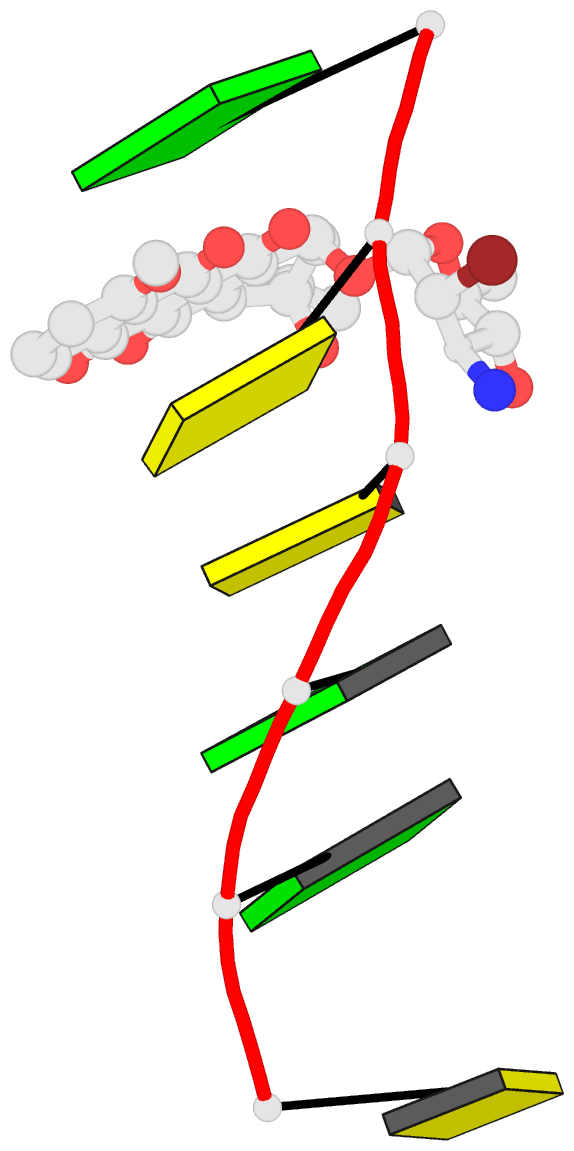
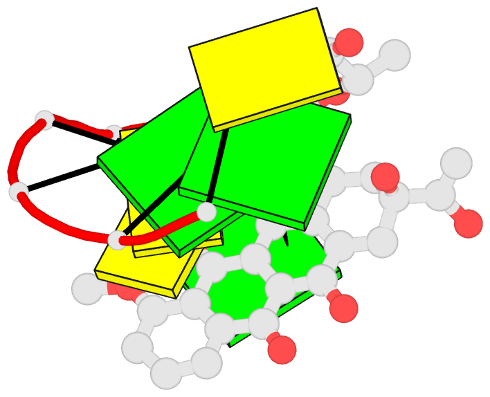
- 278d; DSSR-derived features in text and JSON formats
- Class
- DNA
- Method
- X-ray (1.8 Å)
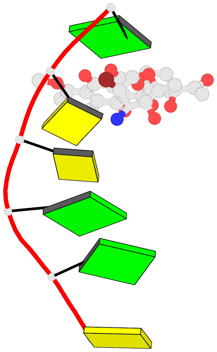
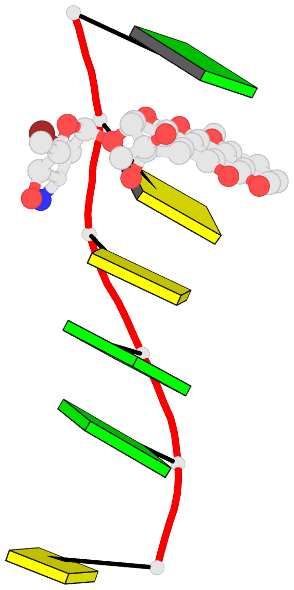
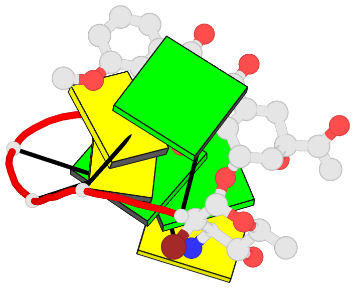
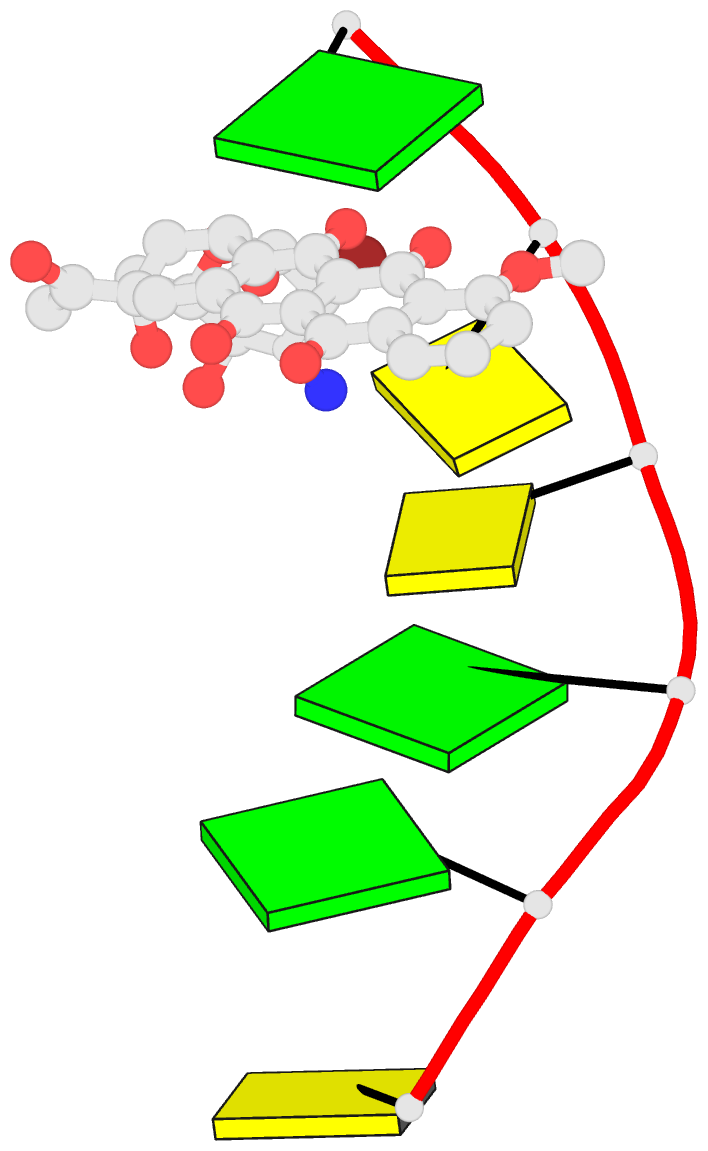
- Summary
- Substitutions at c2' of daunosamine in the anticancer daunorubicin alter its DNA-binding sequence specificity
- Reference
- Gao YG, Priebe W, Wang AH (1996): "Substitutions at C2' of daunosamine in the anticancer drug daunorubicin alter its DNA-binding sequence specificity." Eur.J.Biochem., 240, 331-335. doi: 10.1111/j.1432-1033.1996.0331h.x.
- Abstract
- In the search for new generations of anthracycline drugs, lower cytotoxic side effect and higher activity toward resistant cancer cells are two major goals. A new anthracycline drug, WP401 (2'-bromo-4'-epidaunorubicin, alpha-manno configuration), exhibits promising activity toward multidrug-resistant cells. In contrast, the related compound WP400 (2'-bromo-4'-epidaunorubicin, alpha-gluco configuration), is significantly less cytotoxic. To establish the structural and molecular bases of this observation, we performed X-ray diffraction analyses of the complexes between WP401 and four DNA hexamers CGTACG, CGATCG, CGCGCG, and CGGCCG. Their crystal data (space group P4(1)2(1)2, a = b approximately 2.8 nm, c approximately 5.3 nm) are similar to those of other daunorubicin/doxorubicin complexes. The refined crystal structures at 0.18-nm resolution revealed that two WP401 drug molecules bind to the duplex, with the aglycons intercalated between the CpG steps and their modified daunosamines in the minor groove. The bulky bromine atom at the C2' position caused the daunosamine of the bound WP401 to adopt a different conformation from that of the bound daunorubicin. In the presence of formaldehyde, WP401 formed a covalent adduct with CGGCCG more readily than with CGCGCG. This is the opposite of what is seen for daunorubicin and doxorubicin. Thus modifications at the C2' position of daunosamine modulate the sequence specificity of the formaldehyde-crosslinking reactions between anthracyclines and DNA.